
Identifikasi Alat Diagnostik Untuk Menilai Luka Kronis: Scoping Review
DOI:
https://doi.org/10.24252/asjn.v7i1.62061Keywords:
Diagnostic tool, Chronic wound, non-invasiveAbstract
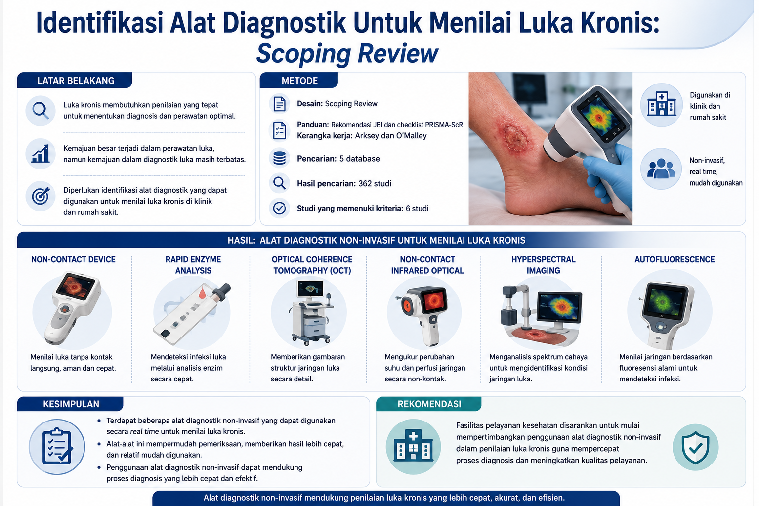
Jutaan orang mengalami luka kronis di seluruh dunia, yang memberikan tekanan yang luar biasa pada sumber daya yang tersedia untuk layanan kesehatan. Meskipun terdapat kemajuan besar dalam perawatan luka, namun hanya sedikit kemajuan dalam diagnostic luka. Dengan melakukan tinjauan pelingkupan, penelitian ini bertujuan untuk mengidentifikasi alat diagnostik yang dapat digunakan untuk menilai luka kronis di Klinik dan Rumah Sakit. Kami mengikuti rekomendasi JBI dan daftar periksa PRISMA-ScR dengan menggunakan Kerangka Kerja Tinjauan Cakupan oleh Arksey dan O'Malley. Pencarian literature dilakukan di lima database, dari 362 studi yang diperiksa, terdapat 6 studi yang memenuhi kriteria untuk ditinjau. Hasil menunjukkan bahwa terdapat beberapa alat diagnostik non-invasif yang dapat digunakan untuk menilai luka kronis secara real time. Termasuk, alat diagnostik non-contact device dan rapid enzim analysis untuk menilai infeksi luka kronis, Optical coherence tomography, non-contact infrared optical, alat diagnostik dengan pencitraan hiperspektral dan Autofluorescence. Tinjauan pelingkupan ini memberikan pemahaman terkait beberapa alat diagnostik non-invasif yang dapat digunakan untuk menilai luka kronis yang mempermudah untuk mengetahui hasil pemeriksaan lebih cepat dan tergolong mudah. Disarankan agar fasilitas pelayanan kesehatan mulai mempertimbangkan penggunaan alat diagnostik non-invasif dalam penilaian luka kronis untuk mempercepat proses diagnosis
Downloads
References
Arafah, M. I. I. M. R. S. (2022). Efektivitas Perawatan Luka Modern dan Konvensional terhadap Proses Penyembuhan Luka Diabetik. Jurnal Ilmiah Mappadising, (Vol 4 No 1 (2022): Jurnal Ilmiah Mappadising Volume 4 Nomor 1 Maret 2022), 1–9. Retrieved from https://ojs.univprima.ac.id/index.php/mappadising/article/view/291/240
Arisandi, D. et al. (2016). Evaluation of validity of the new diabetic foot ulcer assessment scale in Indonesia. Wound Repair and Regeneration, 24(5), 876–884. https://doi.org/https://doi.org/10.1111/wrr.12464
Arksey, H., & O’Malley, L. (2005). Scoping studies: Towards a methodological framework. International Journal of Social Research Methodology: Theory and Practice, 8(1), 19–32. https://doi.org/10.1080/1364557032000119616
Baran, U. et al. (2015). High Resolution Imaging of Acne Lesion Development and Scarring in Human Facial Skin Using OCT-Based Microangiography. 238(December 2014), 231–238. https://doi.org/10.1002/lsm.22339
Bassegoda, A. et al. (2019). Talanta New myeloperoxidase detection system based on enzyme-catalysed oxidative synthesis of a dye for paper-based diagnostic devices. Talanta, 194(August 2018), 469–474. https://doi.org/10.1016/j.talanta.2018.10.065
Bates-Jensen, B. M. et al. (2019). Reliability of the Bates-Jensen wound assessment tool for pressure injury assessment: The pressure ulcer detection study. Wound Repair and Regeneration : Official Publication of the Wound Healing Society [and] the European Tissue Repair Society, 27(4), 386–395. https://doi.org/10.1111/wrr.12714
Blokhuis-Arkes, M. H. E. et al. (2015). Rapid enzyme analysis as a diagnostic tool for wound infection: Comparison between clinical judgment, microbiological analysis, and enzyme analysis. Wound Repair and Regeneration : Official Publication of the Wound Healing Society [and] the European Tissue Repair Society, 23(3), 345–352. https://doi.org/10.1111/wrr.12282
Chin, J. A. et al. (2011). Evaluation of hyperspectral technology for assessing the presence and severity of peripheral artery disease. Journal of Vascular Surgery, 54(6), 1679–1688. https://doi.org/10.1016/j.jvs.2011.06.022
Choi, E. P. H. et al. (2016). Evaluation of the internal and external responsiveness of the Pressure Ulcer Scale for Healing (PUSH) tool for assessing acute and chronic wounds. Journal of Advanced Nursing, 72(5), 1134–1143. https://doi.org/10.1111/jan.12898
Comino-Sanz, I. M. et al. (2023). A Digital Tool for Measuring Healing of Chronic Wounds Treated with an Antioxidant Dressing: A Case Series. International Journal of Environmental Research and Public Health, 20(5). https://doi.org/10.3390/ijerph20054147
DaCosta, R. S. et al. (2015). Point-of-care autofluorescence imaging for real-time sampling and treatment guidance of bioburden in chronic wounds: first-in-human results. PloS One, 10(3), e0116623. https://doi.org/10.1371/journal.pone.0116623
Daeschlein, G. et al. (2017). Hyperspectral imaging as a novel diagnostic tool in microcirculation of wounds. Clinical Hemorheology and Microcirculation, 67(3–4), 467–474. https://doi.org/10.3233/CH-179228
Dahlia et al. (2019). GAMBARAN KARAKTERISTIK LUKA BERDASARKAN SKOR MUNGS DAN ANKLE BRAKHIAL INDEKS ( ABI ) PADA PASIEN DIABETES MELLITUS DENGAN ULKUS DIABETIK. Dunia Keperawatan, 7, 134–141.
Denstedt, M. et al. (2013). Hyperspectral imaging as a diagnostic tool for chronic skin ulcers. Photonic Therapeutics and Diagnostics IX, 8565, 85650N. https://doi.org/10.1117/12.2001087
Frykberg, R. G., & Banks, J. (2015). Challenges in the Treatment of Chronic Wounds. Advances in Wound Care, 4(9), 560–582. https://doi.org/10.1089/wound.2015.0635
Gariani, K. et al. (2013). Diabetic foot infections : state-of-the-art. 1–12.
Godavarty, A et al. (2015). Non-contact optical imaging of healing and non-healing diabetic foot ulcers. 9318, 2–5. https://doi.org/10.1117/12.2080716
Godavarty, Anuradha et al. (2015). Diabetic Wound Imaging Using a Noncontact Near-Infrared Scanner : A Pilot Study. https://doi.org/10.1177/1932296815590828
Greatrex-White, S., & Moxey, H. (2015). Wound assessment tools and nurses’ needs: An evaluation study. International Wound Journal, 12(3), 293–301. https://doi.org/10.1111/iwj.12100
Gupta, S. et al. (2021). Chronic wounds: magnitude, socioeconomic burden and consequences. Wounds Asia, 4(1), 8–14.
Halfens, R. J. G., & Stobberingh, E. E. (2013). Swab Versus Biopsy for the Diagnosis of Chronic Infected Wounds. (May), 211–219.
Holmes, J. et al. (2019). Dynamic Optical Coherence Tomography Is a New Technique for Imaging Skin Around Lower Extremity Wounds. International Journal of Lower Extremity Wounds, 18(1), 65–74. https://doi.org/10.1177/1534734618821015
Hoyo, J. et al. (2022). Rapid Colorimetric Detection of Wound Infection with a Fluidic Paper Device. International Journal of Molecular Sciences, 23(16). https://doi.org/10.3390/ijms23169129
Izatt, J. A. et al. (2015). Theory of Optical Coherence Tomography BT - Optical Coherence Tomography: Technology and Applications (W. Drexler & J. G. Fujimoto, eds.). Cham: Springer International Publishing. https://doi.org/10.1007/978-3-319-06419-2_3
Jayachandran, M. et al. (2016). Critical Review of Noninvasive Optical Technologies for Wound Imaging. Advances in Wound Care, 5(8), 349–359. https://doi.org/10.1089/wound.2015.0678
Joanna Briggs Institute. (2020). JBI Manual for Evidence Synthesis. Retrieved from https://synthesismanual.jbi.global
Kee, K. K. et al. (2019). Risk factor analysis on the healing time and infection rate of diabetic foot ulcers in a referral wound care clinic. 28(1), 5–13.
Kim, D. W. et al. (2016). Plasma treatment effect on angiogenesis in wound healing process evaluated in vivo using angiographic optical coherence tomography Plasma treatment effect on angiogenesis in wound healing process evaluated in vivo using angiographic optical coherence tomo. 233701. https://doi.org/10.1063/1.4967375
Kolluri, R. et al. (2022). An estimate of the economic burden of venous leg ulcers associated with deep venous disease. Vascular Medicine (London, England), 27(1), 63–72. https://doi.org/10.1177/1358863X211028298
Kuck, M. et al. (2014). Evaluation of optical coherence tomography as a non-invasive diagnostic tool in cutaneous wound healing. Skin Research and Technology, 20(1), 1–7. https://doi.org/10.1111/srt.12077
Lei, J. et al. (2018). Assessing the Healing of Venous Leg Ulcers Using a Noncontact Near-Infrared Optical Imaging Approach. Advances in Wound Care, 7(4), 134–143. https://doi.org/10.1089/wound.2017.0745
Lentsck, M. H. et al. (2018). Quality of life related to clinical aspects in people with chronic wound. Revista Da Escola de Enfermagem, 52, 1–9. https://doi.org/10.1590/S1980-220X2017004003384
Li, S. et al. (2020). Imaging in Chronic Wound Diagnostics. Advances in Wound Care, 9(5), 245–263. https://doi.org/10.1089/wound.2019.0967
Li, S. et al. (2021). Diagnostics for Wound Infections. Advances in Wound Care, 10(6), 317–327. https://doi.org/10.1089/wound.2019.1103
Lu, G. et al. (2017). Detection of Head and Neck Cancer in Surgical Specimens Using Quantitative Hyperspectral Imaging. Clinical Cancer Research : An Official Journal of the American Association for Cancer Research, 23(18), 5426–5436. https://doi.org/10.1158/1078-0432.CCR-17-0906
Lu, G., & Fei, B. (2014). Medical hyperspectral imaging: a review. Journal of Biomedical Optics, 19(1), 10901. https://doi.org/10.1117/1.JBO.19.1.010901
McLister, A. et al. (2016). New Developments in Smart Bandage Technologies for Wound Diagnostics. Advanced Materials, 28(27), 5732–5737. https://doi.org/10.1002/adma.201504829
Downloads
Published
How to Cite
Issue
Section
License
Copyright (c) 2026 Nian Yasin, Saldy Yusuf

This work is licensed under a Creative Commons Attribution-NonCommercial-NoDerivatives 4.0 International License.
1.png)
